What Is a Panoramic Dental X‑Ray? Why and How It’s Taken
What Is a Panoramic Dental X‑Ray? Why and How It’s Taken
What Is a Panoramic Dental X‑Ray?
A panoramic dental X‑ray is a special type of dental imaging that captures a single, wide‑angle view of your entire mouth and jaws. This includes:
-
*Upper and lower jaws
-
*All teeth (visible or impacted)
-
*Surrounding bone structures
-
*Jaw joints (TMJ)
-
*Sinuses and nasal area
Because our jaws form a curved arch, this X‑ray transforms that curved shape into a flat image — giving your dentist a broad overview of oral and facial structures at once.
Unlike traditional intra‑oral X‑rays (where a film or sensor is placed inside the mouth), the panoramic X‑ray uses a machine whose arms rotate around your head, making the process much more comfortable and less invasive.
Why Dentists Use Panoramic X‑Rays
Dentists rely on a panoramic X‑ray when they need to examine more than just individual teeth. Common reasons include:
-
*Evaluating impacted or wisdom teeth, seeing their position relative to bone and nerves.
-
*Checking jawbone health, bone abnormalities, cysts, or structural irregularities.
-
*Planning implants, dentures, or major restorative work — to assess bone amount, density, and anatomy.
-
*Preparing for orthodontic (braces) treatment, evaluating jaw alignment, tooth roots, and bone structure.
-
*Detecting infections, tumors, bone lesions, fractures, or sinus‑related dental issues.
-
*Analyzing jaw joints (TMJ) and sinus regions when needed for oral/mouth‑related problems.
Because panoramic X‑rays cover wide anatomical areas, they’re especially valuable before complex dental or surgical treatments — giving a comprehensive baseline.
How a Panoramic X‑Ray Is Taken
Here’s what usually happens during a panoramic dental X‑ray session:
-
Preparation — Patients remove metal objects like jewelry or glasses. A protective apron may be provided.
-
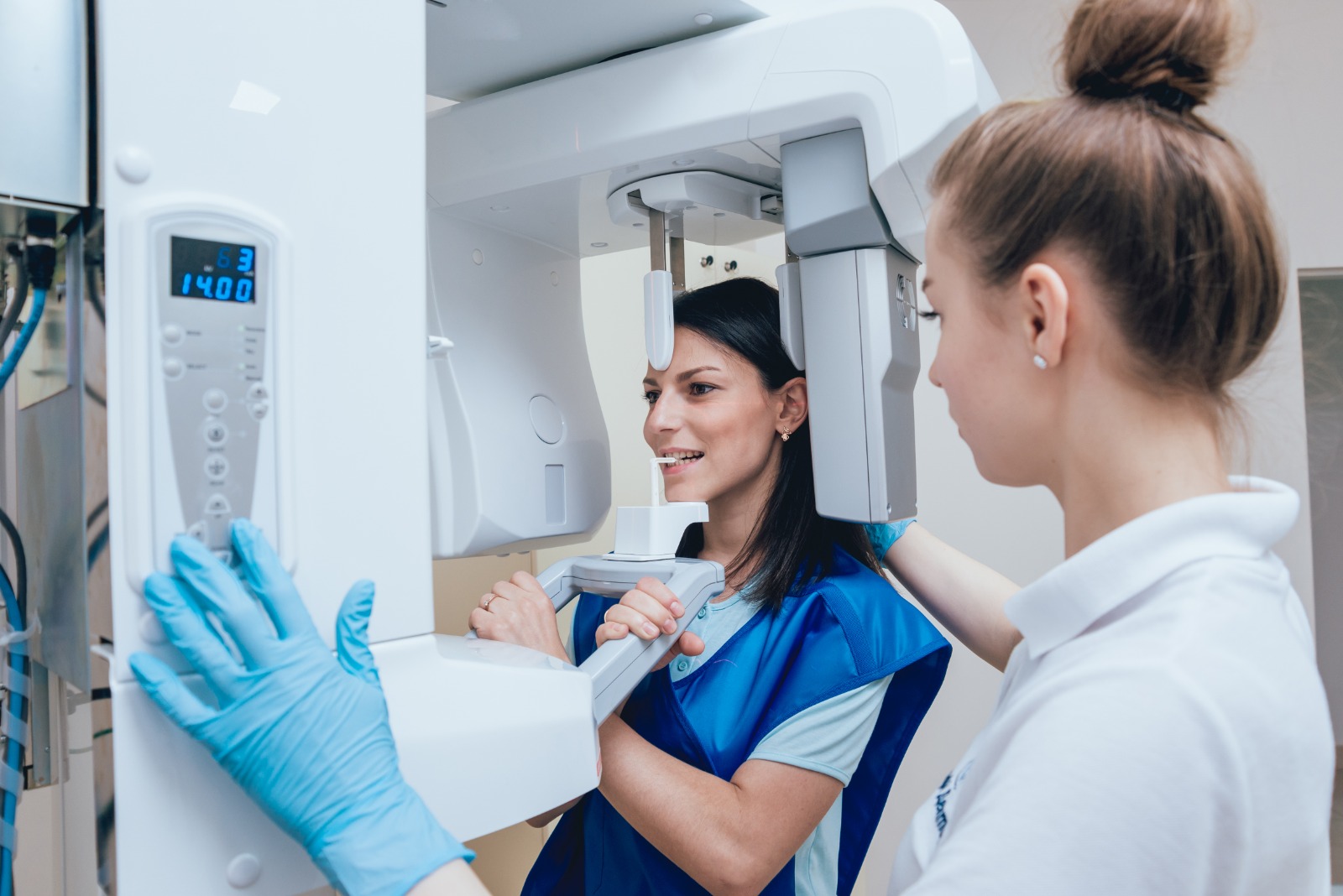
Positioning — You stand or sit in the X‑ray unit; your head is stabilized with supports and often you bite gently on a small plastic piece to align your teeth.
-
Scan — A rotating arm moves around your head while emitting X‑rays; the “film” or sensor is inside the machine, not in your mouth. The scan typically lasts about 10–20 seconds and is painless.
-
Image Generation — A single, panoramic image is produced that shows jaws, teeth, bone structure, sinuses, and nearby anatomical areas. The dentist reviews this image to plan treatments or further diagnostics.
The method is quick, comfortable, and more tolerable for those who might struggle with traditional intra‑oral X‑rays (e.g. strong gag reflex or difficulty opening the mouth).
Advantages & Limitations of Panoramic X‑Rays
Advantages
-
*Gives a full-mouth view — teeth, jaws, bone structure, sinuses and joints — all in one image.
-
*Fast and comfortable — no need for painful or uncomfortable film placement inside the mouth.
-
*Helps detect hidden issues (impacted teeth, bone problems, structural anomalies) that a regular dental exam might miss.
-
*Useful in treatment planning — especially for implants, oral surgery, orthodontics, extractions — anywhere broad anatomical context matters.
Limitations
-
*Lower resolution for small-scale details — fine cavities between teeth, tiny fractures or very early decay may not be visible.
-
*Soft tissues (gums, nerves, muscles) are not clearly visible — the X‑ray primarily shows bones and hard tissues.
-
*Because it flattens a curved 3D structure into 2D, there can be distortions or overlap; precise measurements or diagnoses may require additional intra‑oral X‑rays or 3D imaging (when needed).
In short: panoramic X‑rays are excellent for broad diagnostics and treatment planning, but not ideal for very detailed, close‑up dental issues.
Conclusion — When a Panoramic X‑Ray Makes Sense
If you need a full-picture view of your oral and jaw health — whether for wisdom teeth evaluation, implant planning, orthodontic treatment, jawbone assessment, or sinus/joint analysis — a panoramic dental X‑ray is a powerful, practical tool.
It gives your dentist a comprehensive view with minimal discomfort and helps detect both visible and hidden problems. For many dental and oral‑surgical treatments, it’s often the first essential imaging step.
If your dentist recommends a panoramic X‑ray — it’s usually a smart and beneficial decision for diagnosing and planning effective treatment.